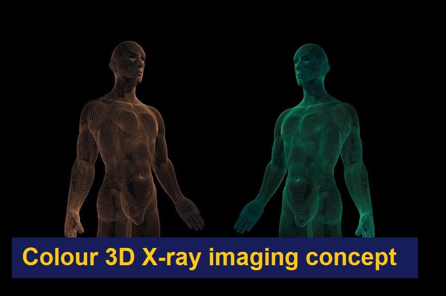
It is heartening to hear about the invention of the colour X-ray that too in a 3D form which can be manipulated to review the anatomical details and diseases of the human body. It looks unusual because we are accustomed to the black and white films of X-ray images. Thanks to the New Zealand-based company MARS Bioimaging that has produced this new technology (Bioscanner) for the detection of diseases. It has been almost 120 years since the discovery of the X-ray by Roentgen, and this new invention will reveal more than mere bone and soft tissue details of the body without cutting open the tissue. These images will definitely improve the efficiency of diagnosis by doctors.
They use sensor chips developed by the European Organisation for Nuclear Research (CERN) which has resulted in the creation of a scanner that produces 3D colour X-Rays of human body tissues. These new impressive images have the ability to clearly define the details of a patient’s bones, lipids and soft tissue as well as other elements such as disease markers. These radiographs have the potential to make accurate diagnosis for different health conditions along with bone damage.
It is a project that took 20 years to complete. It was led by the father-son duo, Phil and Anthony Butler, from the Canterbury and Otago universities respectively. Their scanner is “the first commercially available 3D spectral (multi-energy) scanner to produce in vivo images with anatomic and molecular quantification.”
The new system is based on CERN’s Medipix technology. This is considered to be the most advanced chip today, capable of detecting each individual particle hitting a pixel. “This technology sets the machine apart diagnostically because its small pixels and accurate energy resolution mean that this new imaging tool is able to get images that no other imaging tool can achieve,” explained Phil Butler in the CERN statement. Medipix are particles imaging and detection chips which work in the same way as a camera. Here, a different colour will be assigned to all the detected and counted particles; every individual particle hitting the detector and depending on how many X-rays hit a certain area. It was originally built for the Large Hadron Collider at CERN to track particles but the latest versions of the Medipix chips have since been seen to offer potential in applications outside of high-energy physics.
This 3D scanner is a combination of a strong algorithm and incredible detail. Different tissues, which consist of fat and water, amongst other things, can be imaged and identified by the energy of the X-rays from these different parts. The scanner differentiates bone, muscle, fat, liquids, and all the other materials in the human body. Additional software uses that data to produce full-colour images and that allows a three-dimensional view of the inside of the body. Lately, MARS has been using smaller versions of the scanner to study vascular diseases that cause heart attacks and also cancer related health issues.
The University of Canterbury has come up with a report about the great father and son scientists Phil and Anthony Butler, who had invented the scanner. The first scanning was done on Phil Butler’s ankle and wrist. The machine can detect clearly and illustrate elements such as fat, water, calcium, and, more importantly, disease markers. So, the potential applications in medical imaging are significant.
The researchers have already experimented with using the scanner in the study of ailments such as cancer, bone, joint health, and vascular diseases. “In all of these studies, promising early results suggest that when spectral imaging is routinely used in clinics, it will enable more accurate diagnosis and personalisation of treatment,” said Anthony Butler.